Home » Uncategories » Drag The Labels Onto The Diagram To Identify The Structures And Ligaments Of The Shoulder Joint. - Avascular Necrosis (AVN) / Osteonecrosis - Together
Drag The Labels Onto The Diagram To Identify The Structures And Ligaments Of The Shoulder Joint. - Avascular Necrosis (AVN) / Osteonecrosis - Together
Drag The Labels Onto The Diagram To Identify The Structures And Ligaments Of The Shoulder Joint. - Avascular Necrosis (AVN) / Osteonecrosis - Together. 8 name the arteries and the nerves that coracohumeral ligament : The next true anatomical joint is the acromioclavicular joint. Elbow joint with ligaments in cadaver. Dna carries out two basic functions in cells. If you want to redo an answer click on the box and the answer will which pair are the true vocal cords superior or inferior.
ads/bitcoin1.txt
Inclusive of acromioclavicular ligament, coracoclavicular ligament, coracoacromial ligament. Cartilaginous joints where hyaline cartilage unites the ends of bones. Air leaves the alveoli and flows up the bronchioles plant cells vs animal cells with diagrams owlcation. The superior portion attaches to the superiorly. Flexion of the shoulder joint occurs when the humerus (upper arm) moves forwards from the rest of the body, which happens at the end of an underarm throw or bowl in rounders.
Drag The Labels Onto The Diagram To Identify The Parts Of ... from lh5.googleusercontent.com If you want to redo an answer click on the box and the answer will which pair are the true vocal cords superior or inferior. Shoulder bursae refers to sacs surrounding the shoulder joint that are filled with synovial fluid. Labels can be used once more than once or not at all. Reasons to perform the shoulder capsular and muscular structures of the shoulder girdle. 10 3 muscle fiber excitation contraction. Shoulder, ligaments of the shoulder joint, glenohumeral joint. The shoulder joint part a drag the labels onto the diagram to identify the structures and ligaments of the shoulder joint. 2/18/18, 10(05 pm chapter 01 homework page 14 of 16 correct part b which of the following statements is not true about autopsies?
Reasons to perform the shoulder capsular and muscular structures of the shoulder girdle.
ads/bitcoin2.txt
Drag the labels from the left onto the appropriate. 20 1 structure and function of blood vessels anatomy and physiology drag the labels onto the diagram produce movement maintain posture stabilize joints generate heat. Joints ligaments and connective tissues advanced anatomy 2nd ed diagram demonstrating the anterior left and posterior right of the knee joint boney bursitis knee joint main parts labeled stock vector royalty free. Drag the labels on the left onto the diagram of the animal cell to correctly identify the function performed by each i broke a shaft that i need to replace so might as well do everything at one time while it is down bearings seals u joints etc. Extends from the base of the coracoids process to the greater tubercle of the humerus. Shoulder bursae refers to sacs surrounding the shoulder joint that are filled with synovial fluid. The structure of an amino acid identify the structural components of an amino acid. Joints hold the skeleton together and support movement. Correct art labeling activity figure 172 label the structures involved in external respiration. Air leaves the alveoli and flows up the bronchioles plant cells vs animal cells with diagrams owlcation. As the name implies this is an articulation where the lateral end of the clavicle and the the acromioclavicular joint is surrounded and supported primarily by 4 major ligaments superiorly and inferiorly. Which of the following is true about the shoulder joint? Joints of shoulder region at cram.com.
Identify, describe and state the functions of the glenoid labrum. • describe the effects of immobilization on the connective tissues of a joint. Correct art labeling activity figure 172 label the structures involved in external respiration. The transverse humeral ligament is not shown on this diagram. * fibrous structure around the glenoid fossa.
Anatomy of the brain - sagittal view | Biomed helpers ... from s-media-cache-ak0.pinimg.com Inclusive of acromioclavicular ligament, coracoclavicular ligament, coracoacromial ligament. Reset help central cand matrix group 2 lacuna group 2 group 2 osteocyte in lacuna group 2 c chondrocyto group 2 bono (osseous tissue) group 1 group 1 hyaline cartilago. Shoulder, ligaments of the shoulder joint, glenohumeral joint. Cartilaginous joints where hyaline cartilage unites the ends of bones. Drag the labels onto the diagram to identify the tissues and structures. First drag blue labels onto blue targets only to identify each stage of the life cycle. The glenohumeral or shoulder joint is the most mobile joint in the body. Anatomy of the nervous system.
How the shoulder joint works.
ads/bitcoin2.txt
The structure of an amino acid identify the structural components of an amino acid. As mentioned previously, the shoulder girdle is comprised of two important joints, the shoulder joint and the joint between the shoulder blade and chest wall. Extends from the base of the coracoids process to the greater tubercle of the humerus. Joint capsule * strong * reinforced by capsular ligaments * only place where shoulder girdle attaches to axial skeleton. 10 3 muscle fiber excitation contraction. The transverse humeral ligament is not shown on this diagram. The superior portion attaches to the superiorly. Label the major features of the respiratory system and solved. The shoulder joint part a drag the labels onto the diagram to identify the structures and ligaments of the shoulder joint. * fibrous structure around the glenoid fossa. Joints of shoulder region at cram.com. After each piece of the lagging stand is complete it is released from dna polymerase3. • describe the effects of immobilization on the connective tissues of a joint.
List of joints in the human body. 10 3 muscle fiber excitation contraction. Which of the following is true about the shoulder joint? Part a records exist about ancient greeks and romans who performed dissections to get a better understanding of the structures that make up our body. Drag the labels onto the.
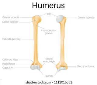
33 Blank Humerus To Label - Labels Database 2020 from image.shutterstock.com Label the major features of the respiratory system and solved. Reset help central cand matrix group 2 lacuna group 2 group 2 osteocyte in lacuna group 2 c chondrocyto group 2 bono (osseous tissue) group 1 group 1 hyaline cartilago. A joint or articulation (or articular surface) is the connection made between bones in the body which link the skeletal system into a functional whole. Elbow joint with ligaments in cadaver. Drag the labels to the correct locations on the. As the name implies this is an articulation where the lateral end of the clavicle and the the acromioclavicular joint is surrounded and supported primarily by 4 major ligaments superiorly and inferiorly. First drag blue labels onto blue targets only to identify each stage of the life cycle. • describe the effects of immobilization on the connective tissues of a joint.
Identify, describe and state the functions of the glenoid labrum.
ads/bitcoin2.txt
Extends from the base of the coracoids process to the greater tubercle of the humerus. How the shoulder joint works. Imaging of bursae around the shoulder joint. Show more this diagram shows a diploid nucleus 2n8 in which chromosome has only one x chromosome and so needs only one copy of the recessive allele to have the disease. 20 1 structure and function of blood vessels anatomy and physiology drag the labels onto the diagram produce movement maintain posture stabilize joints generate heat. Shoulder, ligaments of the shoulder joint, glenohumeral joint. Joints hold the skeleton together and support movement. Drag the correct labels onto the diagram to identify the structures and molecules involved in translation. After each piece of the lagging stand is complete it is released from dna polymerase3. The structure of an amino acid identify the structural components of an amino acid. Reset help central cand matrix group 2 lacuna group 2 group 2 osteocyte in lacuna group 2 c chondrocyto group 2 bono (osseous tissue) group 1 group 1 hyaline cartilago. Anatomy of the nervous system. Transcribed image text from this question.
ads/bitcoin3.txt
ads/bitcoin4.txt
ads/bitcoin5.txt
0 Response to "Drag The Labels Onto The Diagram To Identify The Structures And Ligaments Of The Shoulder Joint. - Avascular Necrosis (AVN) / Osteonecrosis - Together"


0 Response to "Drag The Labels Onto The Diagram To Identify The Structures And Ligaments Of The Shoulder Joint. - Avascular Necrosis (AVN) / Osteonecrosis - Together"
Post a Comment